Product description
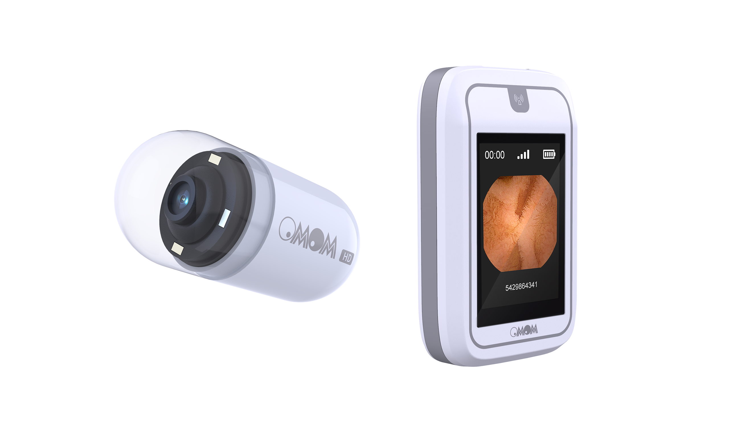
The OMOM HD Capsule Endoscopy System with VUE Smart is a modern, AI-assisted system designed for targeted diagnosis of small bowel diseases.
The system combines a high-resolution imaging capsule, a portable data recorder and the Vue Smart software with integrated AI analysis (SmartScan). The goal is to achieve greater diagnostic precision, significantly reduced reading time and an intuitive, efficient workflow for medical professionals.
Indications
The OMOM HD system is intended for the diagnosis of gastrointestinal tract diseases, particularly in cases of:
• Obscure gastrointestinal bleeding
• Iron deficiency anaemia
• Suspected Crohn’s disease
• Suspected small bowel tumours
• Small bowel polyposis syndromes
• Unexplained malabsorption (e.g. coeliac disease)
• NSAID-associated mucosal injury
• Exclusion of small bowel disease
System Components at a Glance
The OMOM HD Capsule Endoscopy System consists of three main components:
• HD Smart Capsule (OMOM HD)
• Image Recorder
• Vue Smart Software (software platform)
HD Smart Capsule (OMOM HD)
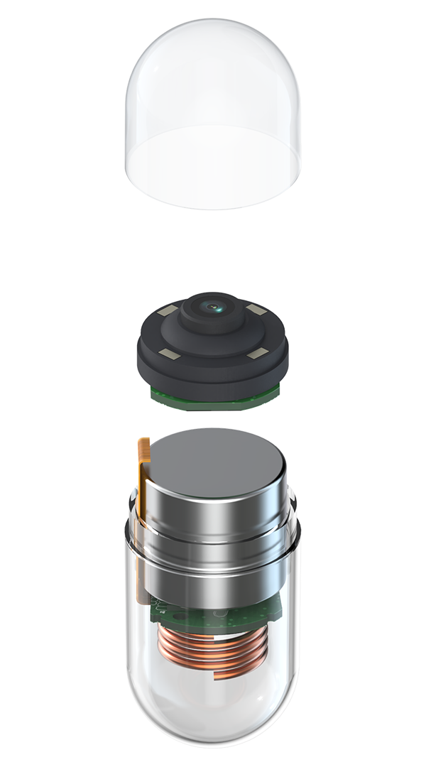
The HD Smart Capsule (OMOM HD) is the central diagnostic element of the system and was specifically developed for reliable and detailed assessment of the small bowel mucosa.
Image Resolution: 512 × 512 pixels
High image resolution is essential for visualising fine mucosal structures. Studies from conventional endoscopy demonstrate that improved resolution significantly increases the detection rate of adenomas and premalignant lesions.
Transferred to capsule endoscopy, this means: the clearer and more detailed the image information, the higher the diagnostic confidence—particularly for subtle, flat or early neoplastic changes.
The 512 × 512 pixel image resolution of the OMOM HD capsule enables precise visualisation of the small bowel mucosa and supports improved assessment of potentially malignant findings.
Size: 25.4 × 11.0 mm
Thanks to its compact design measuring 25.4 × 11.0 mm and a weight of only 3.0 g, the capsule is easy to swallow and well tolerated by patients.
Operating Time: up to 12 hours
Long capsule operating time is a key factor for complete small bowel examination. The combination of energy-efficient image acquisition and adaptive frame rate increases the likelihood of fully visualising the entire small bowel within a single examination. This not only improves diagnostic reach but also reduces the risk of incomplete studies.
Adaptive Frame Rate: 2–10 frames per second (SpeedSense™ technology)
SpeedSense™ technology dynamically adapts the capsule’s frame rate to the individual transit speed within the gastrointestinal tract.
During slow transit, the frame rate is reduced to 2–4 frames per second. This conserves energy and extends the overall operating time of the capsule.
In segments with rapid transit, the frame rate automatically increases to up to 10 frames per second. This minimises the risk of missing small lesions between two frames.
In addition, the adaptive frame rate reduces data volume compared with a constantly high acquisition frequency, resulting in fewer redundant images, more efficient storage and shorter reading times.
Field of View: 172° Ultra-Wide Angle
A wide field of view increases tissue coverage per image and reduces the risk of missing lesions outside the central visual field. Especially in the small bowel, with its complex anatomy and variable folding, an ultra-wide angle helps reliably capture peripheral or partially obscured mucosal areas. Improved surface coverage per image therefore supports a more comprehensive examination of the intestinal lumen.
Image Recorder
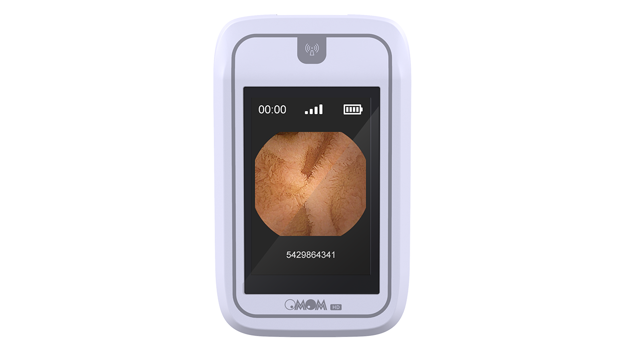
The Image Recorder receives and stores images transmitted by the capsule and enables real-time viewing during the examination. Auto-pairing allows the recorder to connect automatically to the capsule, significantly reducing preparation time.
Preparation and initiation of the examination can be performed directly via the recorder or on a computer. Patient data can be flexibly added and correctly assigned in the Vue Smart workstation before, during or after the examination using the serial number.
After completion of the examination, the image data are available for analysis within approximately 5–7 minutes thanks to fast data download.
Vue Smart Workstation (Software)
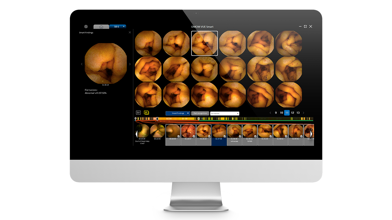
The Vue Smart workstation is the central software platform for data management, analysis and reporting.
Main Functions:
• Patient management
• Capsule–recorder pairing
• Data import from the recorder
• Image analysis and findings documentation
• Report generation and printing
• Data export and import
• SmartScan – AI-assisted lesion detection
AI Function: SmartScan
SmartScan is the integrated AI solution within the Vue Smart software and supports capsule endoscopy reading by automatically prioritising relevant images. The goal is to drastically reduce review time while significantly increasing diagnostic confidence.
Clinical Evidence and Data Basis
SmartScan’s performance is based on an exceptionally broad clinical and data-driven foundation. For development, training and validation, 5,825 small bowel capsule endoscopy examinations from 51 medical centres were used. In total, more than 295 million individual images were incorporated into AI development.
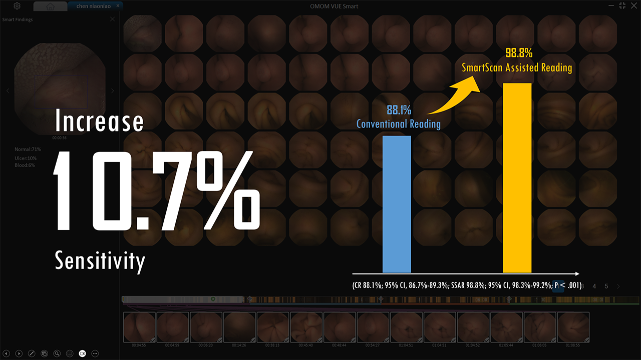
Clinical performance was evaluated in a multicentre comparative study published in JAMA Network Open, directly comparing SmartScan-assisted reading (SSAR) with conventional reading (CR).
The results demonstrate clear clinical benefit:
• +10.7% higher sensitivity in detecting structured small bowel findings
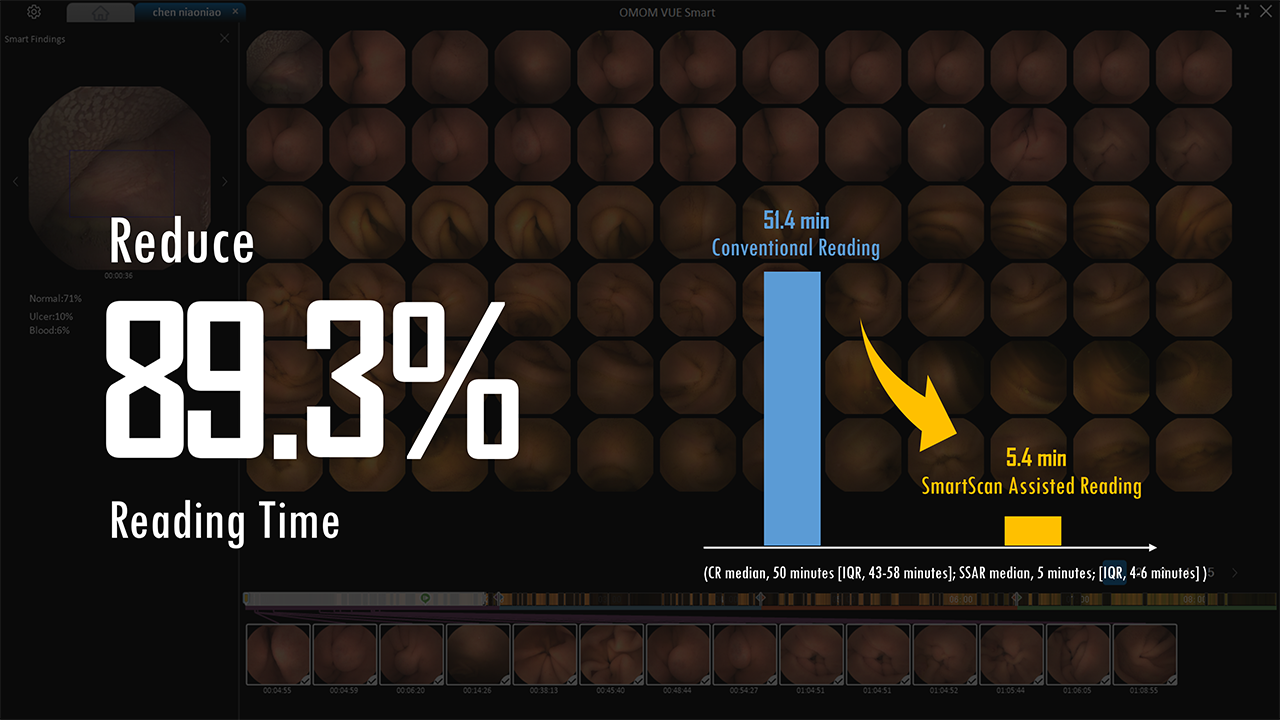
• −89.3% reduction in reading time, decreasing average reading time from approximately 50 minutes to around 5 minutes per examination
• 96% reduction in images requiring review, from approximately 28,000 images to fewer than 800 images per examination
Validation in Clinical Practice
These results were subsequently confirmed under routine clinical conditions in further studies. In a practice-based investigation using OMOM capsules, complete concordance of overall diagnosis was observed between conventional and AI-assisted reading.
At lesion level, SmartScan identified 98.1% of all clinically significant findings, whereas conventional reading detected only 86.2% in direct comparison. In the same study, a further reduction in average reading time to 2–3 minutes per examination was observed.
Continuous development
These consistent results from controlled studies and routine clinical practice formed the basis for the targeted further development of the AI platform. The SmartScan system is continuously being expanded in order to broaden its range of applications and cover additional gastrointestinal findings.
Originally, SmartScan recognised 17 types of structured small bowel findings. Today, SmartScan already identifies 22 different types of gastrointestinal lesions in the stomach and small bowel and has been trained with more than 2.6 million additional images from magnetically controlled capsule endoscopies.
Despite the increased number of detectable lesion types, the number of images requiring review per examination remains below 1,000, with a demonstrably increased detection rate.
Through continuous training with new clinical datasets, SmartScan continues to evolve and supports physicians over the long term with steadily improving diagnostic performance.
SmartScan Modules
SmartView
SmartView significantly reduces reading time by displaying only image sequences classified by the AI as potentially pathological. Instead of reviewing full video sequences with tens of thousands of individual images, readers view a compact, focused video of relevant passages.
This substantially shortens reading time and supports efficient, focused assessment.
SmartFinding
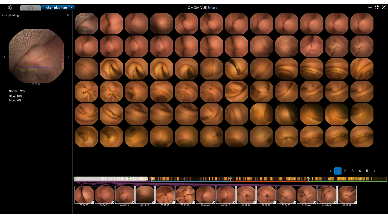
SmartFinding presents detected abnormalities in a structured, page-based view and supports reporting through automatic suggestions for describing clinical findings. The clear layout facilitates systematic evaluation of multiple findings and supports consistent, traceable documentation.
Product description
The OMOM HD Capsule Endoscopy System with VUE Smart is a modern, AI-assisted system designed for targeted diagnosis of small bowel diseases.
The system combines a high-resolution imaging capsule, a portable data recorder and the Vue Smart software with integrated AI analysis (SmartScan). The goal is to achieve greater diagnostic precision, significantly reduced reading time and an intuitive, efficient workflow for medical professionals.
Indications
The OMOM HD system is intended for the diagnosis of gastrointestinal tract diseases, particularly in cases of:
• Obscure gastrointestinal bleeding
• Iron deficiency anaemia
• Suspected Crohn’s disease
• Suspected small bowel tumours
• Small bowel polyposis syndromes
• Unexplained malabsorption (e.g. coeliac disease)
• NSAID-associated mucosal injury
• Exclusion of small bowel disease
System Components at a Glance
The OMOM HD Capsule Endoscopy System consists of three main components:
• HD Smart Capsule (OMOM HD)
• Image Recorder
• Vue Smart Software (software platform)
HD Smart Capsule (OMOM HD)
The HD Smart Capsule (OMOM HD) is the central diagnostic element of the system and was specifically developed for reliable and detailed assessment of the small bowel mucosa.
Image Resolution: 512 × 512 pixels
High image resolution is essential for visualising fine mucosal structures. Studies from conventional endoscopy demonstrate that improved resolution significantly increases the detection rate of adenomas and premalignant lesions.
Transferred to capsule endoscopy, this means: the clearer and more detailed the image information, the higher the diagnostic confidence—particularly for subtle, flat or early neoplastic changes.
The 512 × 512 pixel image resolution of the OMOM HD capsule enables precise visualisation of the small bowel mucosa and supports improved assessment of potentially malignant findings.
Size: 25.4 × 11.0 mm
Thanks to its compact design measuring 25.4 × 11.0 mm and a weight of only 3.0 g, the capsule is easy to swallow and well tolerated by patients.
Operating Time: up to 12 hours
Long capsule operating time is a key factor for complete small bowel examination. The combination of energy-efficient image acquisition and adaptive frame rate increases the likelihood of fully visualising the entire small bowel within a single examination. This not only improves diagnostic reach but also reduces the risk of incomplete studies.
Adaptive Frame Rate: 2–10 frames per second (SpeedSense™ technology)
SpeedSense™ technology dynamically adapts the capsule’s frame rate to the individual transit speed within the gastrointestinal tract.
During slow transit, the frame rate is reduced to 2–4 frames per second. This conserves energy and extends the overall operating time of the capsule.
In segments with rapid transit, the frame rate automatically increases to up to 10 frames per second. This minimises the risk of missing small lesions between two frames.
In addition, the adaptive frame rate reduces data volume compared with a constantly high acquisition frequency, resulting in fewer redundant images, more efficient storage and shorter reading times.
Field of View: 172° Ultra-Wide Angle
A wide field of view increases tissue coverage per image and reduces the risk of missing lesions outside the central visual field. Especially in the small bowel, with its complex anatomy and variable folding, an ultra-wide angle helps reliably capture peripheral or partially obscured mucosal areas. Improved surface coverage per image therefore supports a more comprehensive examination of the intestinal lumen.
Image Recorder
The Image Recorder receives and stores images transmitted by the capsule and enables real-time viewing during the examination. Auto-pairing allows the recorder to connect automatically to the capsule, significantly reducing preparation time.
Preparation and initiation of the examination can be performed directly via the recorder or on a computer. Patient data can be flexibly added and correctly assigned in the Vue Smart workstation before, during or after the examination using the serial number.
After completion of the examination, the image data are available for analysis within approximately 5–7 minutes thanks to fast data download.
Vue Smart Workstation (Software)
The Vue Smart workstation is the central software platform for data management, analysis and reporting.
Main Functions:
• Patient management
• Capsule–recorder pairing
• Data import from the recorder
• Image analysis and findings documentation
• Report generation and printing
• Data export and import
• SmartScan – AI-assisted lesion detection
AI Function: SmartScan
SmartScan is the integrated AI solution within the Vue Smart software and supports capsule endoscopy reading by automatically prioritising relevant images. The goal is to drastically reduce review time while significantly increasing diagnostic confidence.
Clinical Evidence and Data Basis
SmartScan’s performance is based on an exceptionally broad clinical and data-driven foundation. For development, training and validation, 5,825 small bowel capsule endoscopy examinations from 51 medical centres were used. In total, more than 295 million individual images were incorporated into AI development.
Clinical performance was evaluated in a multicentre comparative study published in JAMA Network Open, directly comparing SmartScan-assisted reading (SSAR) with conventional reading (CR).
The results demonstrate clear clinical benefit:
• +10.7% higher sensitivity in detecting structured small bowel findings
• −89.3% reduction in reading time, decreasing average reading time from approximately 50 minutes to around 5 minutes per examination
• 96% reduction in images requiring review, from approximately 28,000 images to fewer than 800 images per examination
Validation in Clinical Practice
These results were subsequently confirmed under routine clinical conditions in further studies. In a practice-based investigation using OMOM capsules, complete concordance of overall diagnosis was observed between conventional and AI-assisted reading.
At lesion level, SmartScan identified 98.1% of all clinically significant findings, whereas conventional reading detected only 86.2% in direct comparison. In the same study, a further reduction in average reading time to 2–3 minutes per examination was observed.
Continuous development
These consistent results from controlled studies and routine clinical practice formed the basis for the targeted further development of the AI platform. The SmartScan system is continuously being expanded in order to broaden its range of applications and cover additional gastrointestinal findings.
Originally, SmartScan recognised 17 types of structured small bowel findings. Today, SmartScan already identifies 22 different types of gastrointestinal lesions in the stomach and small bowel and has been trained with more than 2.6 million additional images from magnetically controlled capsule endoscopies.
Despite the increased number of detectable lesion types, the number of images requiring review per examination remains below 1,000, with a demonstrably increased detection rate.
Through continuous training with new clinical datasets, SmartScan continues to evolve and supports physicians over the long term with steadily improving diagnostic performance.
SmartScan Modules
SmartView
SmartView significantly reduces reading time by displaying only image sequences classified by the AI as potentially pathological. Instead of reviewing full video sequences with tens of thousands of individual images, readers view a compact, focused video of relevant passages.
This substantially shortens reading time and supports efficient, focused assessment.
SmartFinding
SmartFinding presents detected abnormalities in a structured, page-based view and supports reporting through automatic suggestions for describing clinical findings. The clear layout facilitates systematic evaluation of multiple findings and supports consistent, traceable documentation.
Selected products available directly from our warehouse in Zuchwil – we are happy to check additional variants for you
The versions listed below are, depending on the item, either available from stock or upon request.
Looking for a different size or variant? We will be happy to check further options and provide you with individual information on availability and delivery times.
| Article number | Product description | Included in delivery per package | Availability* |
|---|---|---|---|
| CE-5110 | OMOM HD Capsule Endoscope (SC1) | 5 pieces | ✅ In stock in Zuchwil |
| CE-5100 | OMOM HD Capsule Endoscope System (SC-RD1) | 1 piece | ✅ In stock in Zuchwil |
*Orders for in-stock items received by 4:00 p.m. (on business days) are dispatched the same day via A-Post.
Please note: on the day before a public holiday, we are happy to accept orders until 2:00 p.m.
Our consulting team is happy to help you.

Fabio Di Felice
Area Sales Manager for the region Central and South
Certified Medical Technology Consultant shqa
Cantons: BE, SO, TI
+41 79 270 78 51
fdifelice@admedics.com

Anne Rohrbasser
Area Sales Manager for the region West
Certified Medical Technology Consultant
Cantons: FR, GE, JU, NE, VD, VS
+41 79 527 17 63
arohrbasser@admedics.com

Robert Kowalski
Area Sales Manager for the region East
Certified Medical Technology Consultant shqa
Cantons: AI, AR, GL, GR, SG, SH, SZ, TG, UR, ZH, ZG, FL
+41 79 634 59 90
rkowalski@admedics.com

Gina Egli
Area Sales Manager for the region North-Central
Cantons: LU, NW, OW, AG, BL, BS
+41 79 738 60 37
gegli@admedics.com
Certificates and memberships